Imagine waking up one morning and realizing that half of your world has vanished. Not in a dramatic, disappearing act kind of way, but in terms of your field of vision. This is the reality for individuals living with homonymous hemianopsia (HH), a fascinating yet challenging neurological condition that affects how we see the space around us.
If you or someone you know is navigating the complexities of homonymous hemianopsia, you’re not alone. In this comprehensive guide, we will take a look at what homonymous hemianopsia is and why it happens. We will also explore the different features of left and right homonymous hemianopsia as well as incomplete hemianopsia.
Our goal is to help you better understand the condition and break down intricacies in a friendly and easy to understand manner – let’s jump in!
What Exactly is Homonymous Hemianopsia?
Let’s break down the term itself. “Homonymous” means “on the same side,” and “hemianopsia” refers to a loss of vision in half of the visual field. So, homonymous hemianopsia is the loss of vision in the same half of the visual field in both eyes. This is in contrast to bitemporal hemianopsia (also called heteronymous hemianopia), where vision loss occurs on the temporal (outer) side of both eyes.
Think of your visual field as a complete circle surrounding you. Normally, both your eyes work together to capture this entire circle. In order for the brain to integrate information from both eyes and both sides of the visual field, approximately half of the nerve fibers from each eye (the middle portions) cross to the opposite side of the brain, while the other half continue toward the same side of the brain. This crossover occurs at the optic chiasm.
Therefore, information from the right side of what you see goes to the left side of your brain, and information from the left side goes to the right side of your brain. This intricate cross-wiring is what allows us to have a full and integrated visual experience.
However, in individuals with homonymous hemianopsia, this pathway is disrupted, typically due to damage in the brain after the optic chiasm. This means that all of the information from one side of the visual field from both eyes has been lost.
If the visual pathway is disrupted before the optic chiasm, vision loss occurs only in the eye on the side of the damaged nerve, referred to as monocular vision loss. However, damage occurring at the optic chiasm can result in bitemporal hemianopsia, in which the outer portion of the visual field from both eyes is lost.
What Causes Homonymous Hemianopsia? The Common Culprits
Homonymous hemianopsia isn’t a condition that develops on its own. It’s usually a consequence of another neurological event that affects the visual pathways in the brain.
HH frequently arises from vascular injury. In adults, cerebral infarcts (strokes) and intracranial hemorrhages are the most common culprits, accounting for a significant majority of cases. In fact, it is estimated that 52-70% of cases of hemianopsia result from stroke, with 8-10% of all stroke survivors experiencing homonymous hemianopsia permanently.
Other contributing factors include:
- Traumatic Brain Injury (TBI): accounts for 14% of homonymous hemianopsia cases
- Tumors: Particularly prevalent in pediatric cases, responsible for 11% of HH cases
- Iatrogenic Events: Injury resulting from medical treatment, such as neurosurgical procedures or radiation necrosis.
- Demyelinating Disorders: Such as multiple sclerosis (MS).
- Neurologic Diseases: Including Alzheimer’s disease and Creutzfeldt-Jakob disease.
- Vascular Malformations: Like arteriovenous malformations.
- Infections: Such as brain abscesses or toxoplasmosis.
- Inflammatory Diseases: Beyond MS, like neuromyelitis optica.
- Metabolic Diseases: Such as MELAS (mitochondrial encephalomyopathy, lactic acidosis, and stroke-like episodes).
- Seizures or Epilepsy: occipital, temporal or parietal lobe seizures may result in temporary homonymous hemianopsia
- Migraines: can cause temporary homonymous hemianopsia
- Transient Ischemic Attacks (TIAs): may also cause temporary HH with spontaneous recovery
- Nonketotic Hyperglycemia: can resolve with proper blood sugar management
The specific location of the brain lesion dictates the type and extent of the HH. Studies have shown that the most common lesion locations are the occipital lobe, followed by the optic radiation, optic tract, and less frequently, the lateral geniculate nucleus (LGN).
The location and extent of the brain injury will determine which side of the visual field is affected and the severity of the vision loss.
Understanding the Impact: Common Symptoms and Features of Homonymous Hemianopsia
Individuals with homonymous hemianopsia experience a loss of vision in either the right or left half of their visual field in both eyes. This can manifest in various ways:
- Complete Hemianopsia: Loss of the entire half of the visual field on the affected side, including the central macular vision.
- Partial or Incomplete Hemianopsia: Vision loss that doesn’t encompass the entire half of the visual field. This includes:
- Homonymous Quadrantanopia: Loss of one-quarter of the visual field (either upper or lower, on the same side in both eyes). Damage to the upper or lower banks of the visual cortex or specific parts of the optic radiation can cause this.
- HH with Macular Sparing: Retention of central vision despite the hemianopic defect. This is often seen in occipital lobe strokes due to the dual blood supply to the macular region.
- Homonymous Scotomatous Defects: Rare, localized blind spots within the central 30 degrees of vision, respecting the vertical midline; this is somewhat of a reversal of HH with macular sparing.
- Homonymous Sectoranopia: A rare, wedge-shaped loss of vision, often linked to LGN involvement.
- Temporal Crescent Sparing: Loss of the outermost peripheral vision on one side only, resulting from anterior occipital lobe damage on the side of the brain opposite from the eye with vision loss.
Patients with HH may initially report difficulties such as bumping into objects on their blind side, struggling with reading (especially tracking lines of text), and challenges with navigation and driving.
Understanding Left, Right Homonymous Hemianopsia and the Concept of Contralateral
Now, let’s take a look at some of the specifics in how homonymous hemianopsia can manifest. Specifically, we will look at left and right homonymous hemianopsia and the concept of contralateral hemianopsia.
1. Right Homonymous Hemianopsia
Imagine looking straight ahead. If you have right homonymous hemianopsia, you will have lost vision in the right half of your visual field in both eyes. This means you can see things to your left, but everything on your right side will be out of view.
Think about everyday scenarios:
- Walking
- Reading
- Driving
- Eating
With right homonymous hemianopsia, each of these everyday scenarios become significantly more difficult. Reading can be particularly challenging with right HH when trying to read from the left to the right, as it is continually challenging to see the next word.
Right homonymous hemianopsia is typically caused by damage to the left side of the brain, specifically the occipital lobe or the visual pathways leading to it. Remember the cross-wiring described above – the left side of the brain processes information from the right visual field.
2. Left Homonymous Hemianopsia
Conversely, left homonymous hemianopsia involves the loss of vision in the left half of the visual field in both eyes. In other words, you’ll be able to see things on your right, but everything to your left will be invisible.
Left homonymous hemianopsia is usually caused by damage to the right side of the brain, affecting the occipital lobe or the visual pathways leading there. The right side of the brain processes information from the left visual field.
3. Contralateral: The Key Concept
The terms “left” and “right” homonymous hemianopsia inherently involve the concept of contralateral. “Contra” means “opposite,” and “lateral” refers to “side.” In the context of the visual pathways, the brain processes visual information from the opposite side of the visual field.
Therefore:
- Damage to the left side of the brain results in a visual field defect on the right side (right homonymous hemianopsia).
- Damage to the right side of the brain results in a visual field defect on the left side (left homonymous hemianopsia).
Understanding this contralateral relationship is crucial for patients and healthcare professionals to understand the location of the brain injury that caused the vision loss.
Living with Homonymous Hemianopsia: How To Adapt to Challenges in Daily Life
Living with homonymous hemianopsia presents a unique set of challenges that can significantly impact daily life. The loss of half of the visual field can affect:
- Mobility and Navigation
- Reading and Writing
- Grooming and Self Care
- Driving
- Social Interactions
- Work and Hobbies
However, it’s important to emphasize that individuals with homonymous hemianopsia can learn to adapt and lead fulfilling lives. Let’s take a look at several rehabilitation strategies and assistive devices that play a crucial role in this process.
Navigating the Challenges: Rehabilitation and Compensation Strategies for Homonymous Hemianopsia
While lost vision in HH often cannot be restored, various strategies can help individuals adapt and improve their daily functioning.
These include:
- Prisms: Special lenses that can shift the visual field towards the blind side, increasing awareness of objects in that area. Peli prisms, placed in the upper and lower parts of the spectacle lens on the side of the HH, are a common type.
- Visual Scanning Training: Therapy focused on teaching conscious and systematic eye and head movements to compensate for the lost visual field, improving detection of objects on the affected side.
- Exploration Therapies: Techniques aimed at enhancing awareness of the visual field loss and improving visual search strategies.
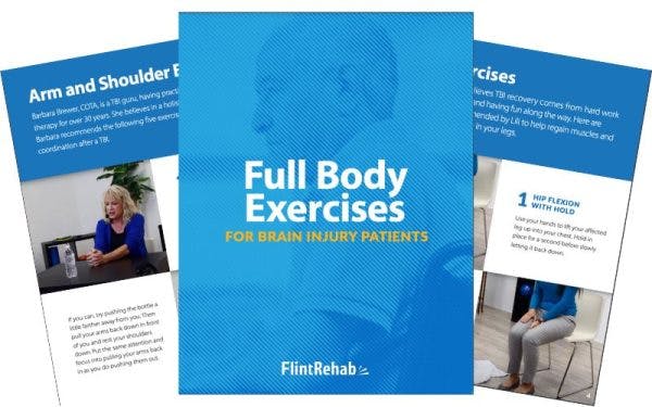
- Occupational Therapy: Helps individuals adapt to daily activities such as dressing, eating, and navigating their environment.
- Orientation and Mobility Training: Specialists teach safe and effective techniques for moving around indoors and outdoors.
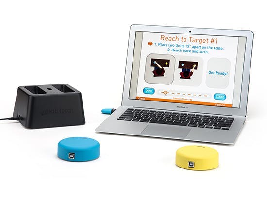
- Assistive Technology: Computer software, screen readers, and navigation apps can aid with reading, writing, and mobility.
- Psychological Support: Addressing the emotional impact of vision loss and providing coping strategies is crucial.
Can You Improve Homonymous Hemianopsia?
Spontaneous improvement of HH can occur, particularly in the initial months after the injury, especially following a stroke. Studies suggest that 50-60% of individuals may experience some visual field recovery within the first month, with most improvement occurring within three to six months. Complete spontaneous recovery is less common, but may occur in 8-12% of those with homonymous hemianopsia.
Spontaneous recovery after six months is less common. At that point, improvements may also be linked to the resolution of underlying medical conditions.
Potential Complications of Homonymous Hemianopsia
Beyond the visual field deficit itself, individuals with HH may experience:
- Increased Risk of Falls and Injuries: Due to reduced awareness of their surroundings.
- Difficulties with Mobility and Navigation: Making everyday tasks like walking and crossing the street challenging.
- Reading and Writing Difficulties: Affecting communication and daily tasks.
- Driving Limitations: Often making driving unsafe and legally restricted in many areas.
- Psychological Impact: Including feelings of disorientation, anxiety, and depression.
Living Fully with Half the World
While the diagnosis of homonymous hemianopsia can be life-altering, it doesn’t define a person’s potential. With dedicated rehabilitation, adaptive strategies, and a strong support system, individuals can learn to navigate their world effectively and participate fully in life. By understanding the nuances of left and right homonymous hemianopsia, as well as incomplete hemianopsia, we can better support those living with this condition and empower them to see their world in new and meaningful ways.
If you or someone you know is experiencing vision loss, it’s crucial to seek professional medical advice promptly. Early diagnosis and intervention are key to maximizing adaptation and improving quality of life. Remember, even with half the visual field, a whole and fulfilling life is still within reach.